Neural Stem Cell Research at Stockton
Summer 2018 Issue

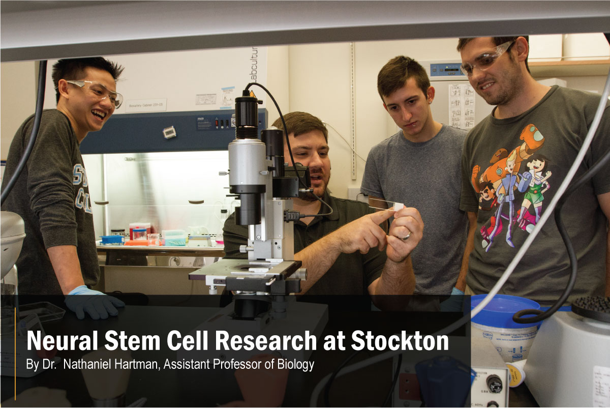
Dr. Nathanial Hartman, Assistant Professor of Biology, with Biology research students
(left to right) Doan Tran, Biology, Physical Therapy
concentration '14, Jeffrey Mercurio, Biology, Behaviorial Neuroscience concentration,
and Michael Alex Finger, Biochemistry '15. (Photo credit: Susan Allen)

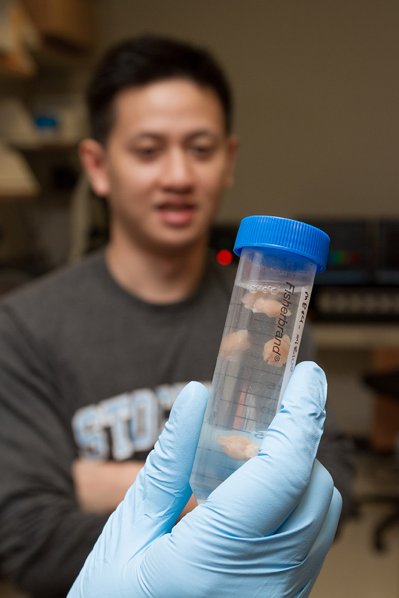
(Foreground) Biology-Physical Therapy alumnus, Doan Tran (background) looks at tube with preserved mouse brains. (Photo credit: Susan Allen)
In the human brain, there are hundreds of billions of highly specialized cells all derived from neural stem cells. In this way, neural stem cells are the foundation upon which the nervous system grows and develops. Each neural stem cell can produce neurons and non-neural cells known as glial cells. There is a multitude of neuron types, with each neuron type differing in how they connect and communicate with other cells. Understanding how neural stem cells give rise to this diverse pool of neurons is critical to understanding human disorders and disease.
Neurodevelopmental disorders arise from improper development in the central nervous system, usually due to dysfunction in neural stem cell behavior. Autism spectrum disorder is a particularly prevalent example where patients exhibit problems in social interaction and repetitive behaviors. The causes of autism are still unclear, stemming from genetic risk factors, prenatal and perinatal factors, and environmental factors. Recent studies have demonstrated alterations in the numbers of neurons produced in certain areas of the brain and dramatic changes to the structure of neurons in patients with autism spectrum disorder.
The best science education requires that undergraduate students get into the laboratory and gain research experience.
In our laboratory, we study how specific molecular pathways converge and alter neural stem cell behavior*. One protein of particular interest is the mammalian target of rapamycin (mTOR). Our lab and others have observed that increased activation of the mTOR pathway in neural stem cells leads to an overproduction of neurons, much larger cells and misplaced neurons. These results mirror what occurs in patients with Tuberous Sclerosis, a genetic disorder with a high comorbidity with autism spectrum. In understanding how mTOR affects neural stem cell behavior, we will open new ideas for treatment and therapy and gain a deeper appreciation for the development of the most complex organ in the known universe, the human brain.
The path from neural stem cell to neuron is complex and tightly regulated. Proper signals instruct the cell to divide and give rise to daughter cells. These new daughter cells must then manage to migrate to the correct location and mature, sending out large and dynamic thin extensions that connect to hundreds and thousands of other cells.
Even slight perturbations can cause extensive problems in the development of neural stem cells into functional neurons. We study how the mTOR pathway is involved in every step of neurogenesis, the creation of new neurons, in the postnatal brain, from initial cell division to final integration in the neural circuit.
There is ongoing neurogenesis in the postnatal brain in all mammal species studied. Neural stem cells that line the fluid-filled ventricles give rise to daughter cells that migrate long distances forward into the olfactory bulb in order to become mature neurons. To study how mTOR affects these processes, we deliver plasmids that can alter gene expression and function to the neural stem cells around the ventricles. We also use these plasmids to express fluorescent proteins in order to track the cells we target. Using these techniques, we can track migration aberrations, changes in cell proliferation rates, and alterations to neuron morphology.

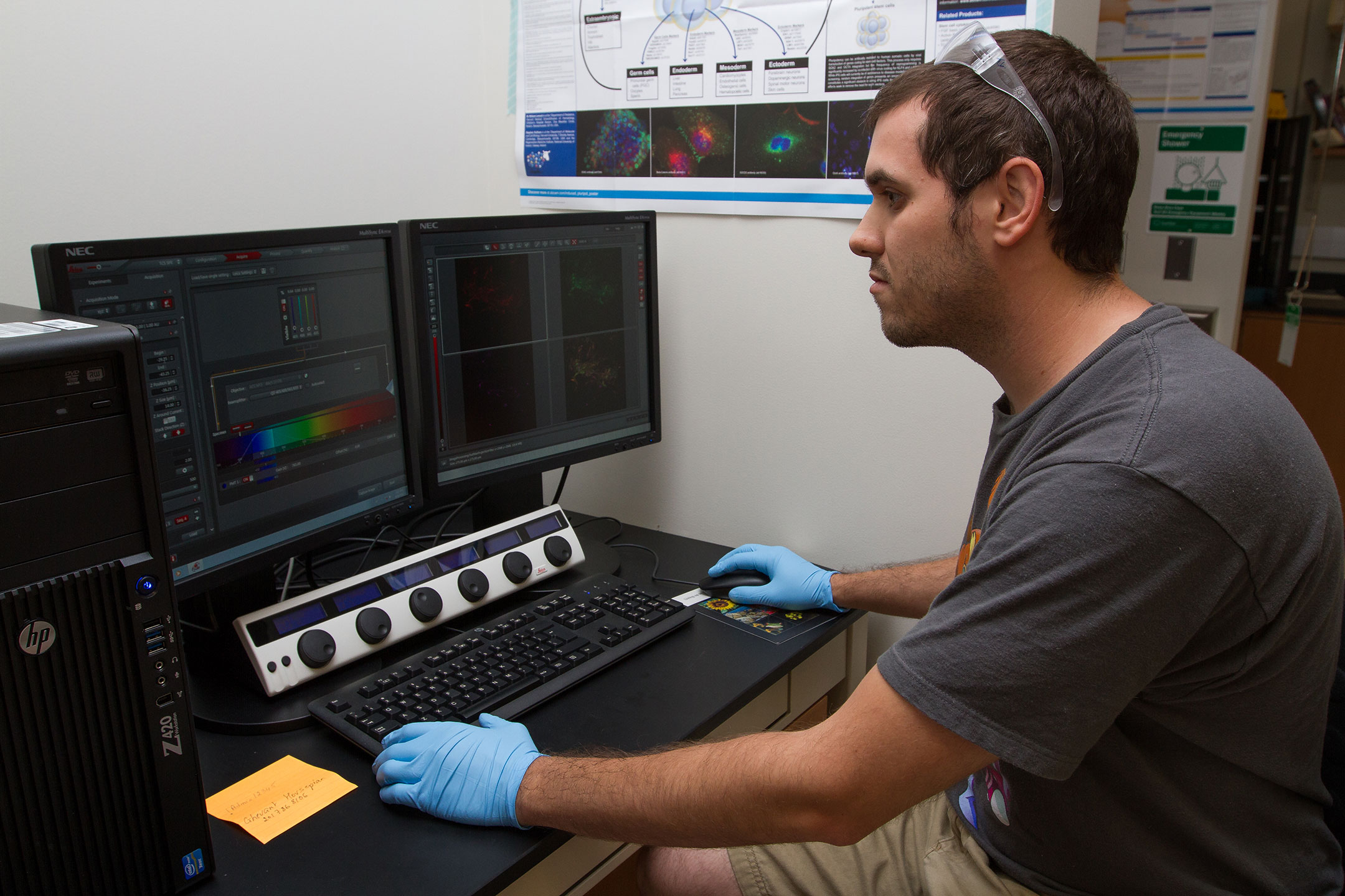
Michael Alex Finger, Biochemistry '15 , uses the confocal microscope to identify neural stem cells in the mouse brain. (Photo credit: Susan Allen)
The best science education requires that undergraduate students get into the laboratory and gain research experience. I have been fortunate to mentor truly amazing students over the past four years. Stockton undergraduates from my lab have completed guided research projects on neurogenesis in the laboratory using these techniques and others. Students have learned to stain cells, measure and analyze neural morphology, run protein analysis and gene expression analysis. Students in the lab have presented their work at Stockton events, regional symposia and international conferences. Their research experiences in the laboratory has propelled them into Ph.D. and M.D. programs, in-vitro fertilization clinics and pharmaceutical companies.
*This research is being funded by the National Institutes of Health (NIH)


